Diagnostic imaging, video endoscopy and pathology
The Equine Centre offers a range of Computed Radiography & Tomography (CT, CAT Scan), Ultrasonography, Scintigraphy, MRI, and Pathology
Computed radiography and digital radiography

The Equine Centre uses both computed radiography and digital radiography systems to obtain high-quality x-rays of all parts of the horse. We have two roof-mounted x-ray tubes and a powerful generator that allows images to be obtained even in large horses. Most radiographs can be taken standing in sedated horses, including radiographs of the shoulder and thorax (chest).
Standing Computed Tomography (CT, CAT scan)
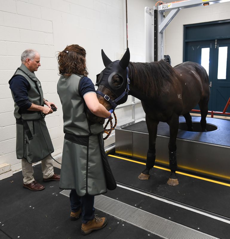
The Equine Centre was the first equine hospital in Australia to have a dedicated CT system for imaging horses standing, under sedation, without the need for anaesthesia. It enables horses to be imaged very rapidly (scan time is approximately 30 seconds). It is capable of imaging both forelimbs from the carpus down to the foot, both hindlimbs from the hock to the foot, as well as the head and upper neck.
CT scanning uses x-rays to create a three-dimensional image of the area of interest. This greatly increases the information obtained and is therefore far more sensitive for identifying both bony and soft tissue lesions when compared with conventional (2D) x-rays. We are also able to image the whole body of foals, which has been extremely useful in identifying subtle changes in sick foals and allowing prompt intervention.
CT is the ideal technique for imaging the head and evaluating animals for diseases of the teeth, sinuses, and nasal cavity. CT can also be extremely useful for assessing bone and soft tissue injuries in the lower limbs and we will often perform a CT for surgical planning or to obtain more information on a condition prior to surgery.
Ultrasonography
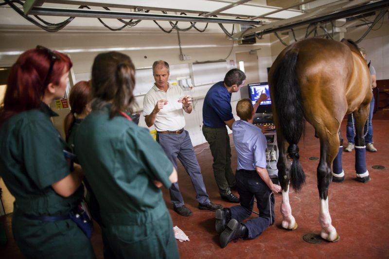
Ultrasound is used primarily for imaging soft tissues, although it is also useful for bone surfaces. It therefore combines well with x-rays for investigation of limb problems. It can be used to investigate bone problems in areas where obtaining good-quality x-rays is problematic, such as the pelvis and spine.
Scintigraphy (Bone scan)


Bone scanning is the most sensitive technique for detecting bone injury available for horses. It involves the injection of a radiopharmaceutical that is taken up by areas of bone with increased turnover. A large camera (gamma camera) is used to image the horse and detect ‘hot spots’ where there is injury.
The Equine Centre Scintigraphy Unit has a proud history over more than 18 years of producing extremely high-quality images. Our qualified and very experienced nuclear medicine staff keep this sophisticated and sensitive equipment running smoothly and accurately. Over the years, we have accumulated a vast database of images with which we can compare each new case. Our current scintigraphy system, the German-made Equine Scanner H.R. Scintron, provides more sophisticated, efficient, and user-friendly scans, with improved comfort and protection to equine patients.
We are able to perform scans on all areas of the body, from the feet to the head. Scintigraphy is especially useful for cases where the source of the lameness is difficult to localise, where the site of lameness has been identified but there are no changes on radiographs, where multiple sites of lameness are suspected, and for problems of the spine, pelvis, and sacroiliac regions. The horse is usually admitted the day before the scan and, depending on results, is able to go home 24 hours after scintigraphy.
Magnetic Resonance Imaging (MRI)
The Equine Centre has the only ultra-high-field MRI scanner in Australia for horses. MRI is the most advanced imaging technique currently available. It allows three-dimensional imaging of both soft tissue and bone, all at once. For horses, it is most valuable for imaging problems within the foot, because ultrasound imaging in this region is limited by the difficulty of penetrating the hoof.
Many horses have lameness that can be localised to the foot with nerve blocks, but radiographs are normal or show changes for which the significance is unknown. MRI is extremely valuable in these cases as the bones can be imaged in far greater detail, as well as the soft tissues. Common problems that occur within the foot that require MRI for a diagnosis are deep digital flexor tendon injuries, injuries to the collateral ligaments of the coffin joint, problems with the ligaments of the navicular bone, and navicular bone injuries themselves.
MRI systems are divided into low-field, high-field and ultra-high-field systems, based on the strength of the magnet. In general, the stronger the magnet (higher field), the more detailed the image quality and the larger the area that can be imaged. Although low-field systems allow imaging while standing and sedated, the disadvantage is that subtle changes may be more difficult to identify due to the smaller areas that can be scanned, the lower resolution, and problems with movement. High-field systems require general anaesthesia, but can image larger areas and provide better resolution, which may lead to more effective diagnosis.
The ultra-high-field system at the Equine Centre can image the foot, pastern and fetlock, and cannon area of most larger-breed adult horses.